Unveiling the Hidden: A Comprehensive Look at X-Ray Machine Images
Related Articles: Unveiling the Hidden: A Comprehensive Look at X-Ray Machine Images
Introduction
With enthusiasm, let’s navigate through the intriguing topic related to Unveiling the Hidden: A Comprehensive Look at X-Ray Machine Images. Let’s weave interesting information and offer fresh perspectives to the readers.
Table of Content
Unveiling the Hidden: A Comprehensive Look at X-Ray Machine Images
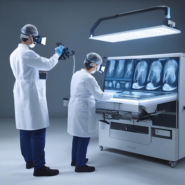
X-ray machines, with their ability to penetrate the human body and reveal its internal structures, have revolutionized medical diagnosis and treatment. The images they produce, known as radiographs, provide invaluable insights into the state of bones, organs, and tissues, aiding in the detection and management of a wide range of conditions.
Understanding the Science Behind X-Ray Images
X-rays are a form of electromagnetic radiation with wavelengths shorter than visible light. When directed at the body, these rays pass through soft tissues relatively easily, while being absorbed to a greater extent by denser materials like bone. This differential absorption creates a shadow image on a detector, showcasing the skeletal framework and any abnormalities within.
Types of X-Ray Machines and their Applications
The world of X-ray technology encompasses a variety of machines tailored to specific medical needs:
- General Purpose X-Ray Machines: These versatile units are commonly found in hospitals and clinics, used for a wide range of examinations including chest X-rays, dental X-rays, and extremity imaging.
- Fluoroscopy Machines: These machines produce real-time images, allowing physicians to observe the movement of internal structures, often used in procedures like barium swallow studies and angiograms.
- Computed Tomography (CT) Scanners: Utilizing a rotating X-ray source and detector, CT scanners generate detailed cross-sectional images of the body, providing a more comprehensive view than traditional X-rays.
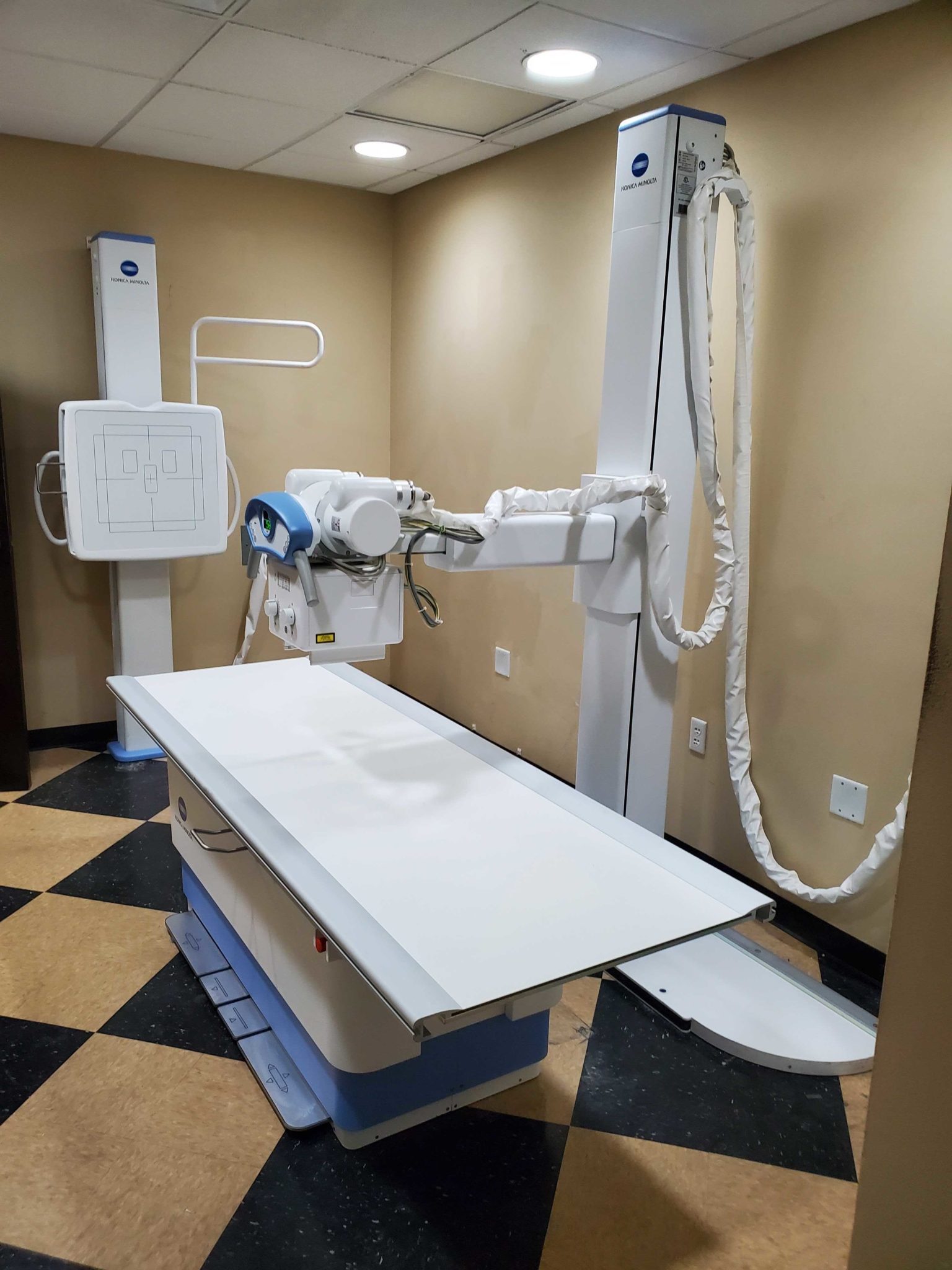
- Digital Radiography (DR) Machines: These systems convert X-ray signals into digital images, offering advantages like faster processing, enhanced image quality, and reduced radiation exposure compared to traditional film-based methods.
The Significance of X-Ray Images in Medical Diagnosis
X-ray images play a crucial role in various medical specialties, providing essential information for:
- Fractures and Dislocations: X-rays clearly depict broken bones and displaced joints, aiding in diagnosis and guiding treatment.
- Infections and Inflammation: X-ray images can reveal signs of infections like pneumonia in the lungs or arthritis in the joints, allowing for timely intervention.
- Tumors and Masses: While not always definitive, X-rays can detect abnormal growths and masses, prompting further investigation with other imaging techniques.
- Foreign Objects: X-rays can locate foreign objects lodged within the body, facilitating their removal.
- Cardiovascular Conditions: X-rays can help assess heart size, detect fluid buildup around the heart, and reveal signs of heart disease.
- Dental Health: Dental X-rays are essential for diagnosing cavities, root canals, and other dental issues.
Beyond Diagnosis: The Role of X-Ray Images in Treatment
X-ray images are not merely diagnostic tools; they also guide treatment strategies:
- Orthopedic Procedures: X-rays are crucial for planning and monitoring surgical procedures involving bones and joints.
- Radiation Therapy: X-rays are used in radiation therapy to target and destroy cancerous cells.
- Interventional Procedures: X-ray guidance allows physicians to perform minimally invasive procedures like biopsies and stent placements.
Benefits of X-Ray Images
The use of X-ray images offers numerous benefits to patients:
- Early Detection: X-rays can detect conditions in their early stages, allowing for more effective treatment.
- Non-Invasive: Most X-ray examinations are non-invasive, avoiding the risks associated with surgery.
- Affordable: X-rays are generally a cost-effective diagnostic tool compared to other imaging techniques.
- Wide Availability: X-ray machines are readily available in most healthcare facilities, making them easily accessible.
Minimizing Risks Associated with X-Ray Exposure
While X-rays are generally safe, excessive exposure can pose risks. Modern X-ray machines employ techniques like digital radiography and radiation shielding to minimize exposure. Healthcare professionals are trained to use X-ray machines responsibly and ensure patient safety.
FAQs about X-Ray Images
Q: How safe are X-rays?
A: X-rays are generally safe when used appropriately. Modern machines and techniques minimize radiation exposure, and healthcare professionals adhere to strict safety protocols.
Q: What are the risks associated with X-rays?
A: While rare, excessive exposure to X-rays can increase the risk of developing cancer. However, the benefits of X-ray diagnosis typically outweigh these risks.
Q: How often can I have an X-ray?
A: The frequency of X-ray examinations depends on individual medical needs and the specific type of X-ray. Healthcare professionals determine the appropriate frequency based on clinical assessment.
Q: What should I do before an X-ray?
A: You may be asked to remove jewelry and metal objects before an X-ray. Inform your healthcare provider about any potential pregnancy or medical conditions.
Q: What should I do after an X-ray?
A: You can typically resume your normal activities after an X-ray. If you experience any unusual symptoms, consult with your healthcare provider.
Tips for Understanding X-Ray Images
- Consult with a Healthcare Professional: Always discuss X-ray results with a qualified physician for accurate interpretation and diagnosis.
- Observe the Image Carefully: Look for any abnormalities, such as fractures, masses, or fluid buildup.
- Pay Attention to Labels and Markers: These indicate the patient’s identity, the date of the examination, and the body part imaged.
- Consider the Context: The image should be interpreted in conjunction with the patient’s medical history and clinical symptoms.
Conclusion
X-ray images have become an indispensable tool in modern medicine, providing valuable insights into the human body and aiding in the diagnosis and treatment of a wide range of conditions. By understanding the science behind these images, their applications, and the associated risks and benefits, individuals can better appreciate their significance in maintaining health and well-being.


Closure
Thus, we hope this article has provided valuable insights into Unveiling the Hidden: A Comprehensive Look at X-Ray Machine Images. We hope you find this article informative and beneficial. See you in our next article!